
Mole Examination
The beautiful thing about moles, especially on the face, is that they add more character to our features. Some moles run in the family which can often look the same and are in the same location in the same way as birthmarks. If you have a history of skin cancer linked to moles, it is important that you get a mole examination. It's a good idea to be proactive and keep an eye out for dangerous moles that have the potential to be cancerous now or in the near future.
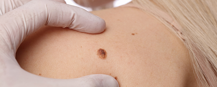
If you notice changes in a mole's colour or appearance, you should have a mole examination. It becomes more urgent if the moles bleed, ooze, itch, appear scaly or become tender or painful. Dr Ayanda Motau will look for any suspicious changes in your moles when you have a skin examination. Your age, sex and when the mole appeared will be taken into consideration. Melanoma is the most common cancer in women ages 25 to 29. New moles in people over the age of 30 are often suspicious.
Possible skin cancer signs in moles:
- An asymmetrical mole that was previously symmetrical.
- Blurred and ridged outer edges.
- The diameter on the mole is larger than normal.
- The mole is continuously changing shape and size.
- The mole has a variety of colours ranging from browns, blue, black and reds.
It is vital that you keep track of the moles that you have. It is possible to develop moles over time which could be mistaken for freckles but could be telling you a different story. Your skin is a large organ and is often the first organ to signal irregularities, health threats and bodily changes. For this reason, we must be attentive to multiple moles the appear out of nowhere or start to give problems, even if they seem like minor changes throughout your lifetime.
Mole Mapping
Using MoleMax® HD detailed imaging of your melanocytic nevi (moles) and other skin lesions can be mapped over your whole body.
MoleMax® is the original Computerised Digital Epiluminecence developed in the 1990s. Since then it has undergone numerous updates. The current version allows for up to 90x magnification of skin lesions.
The entire procedure takes about 30 to 45 minutes depending on the number of moles you have. Detailed imagery allows for more accurate diagnosis of the skin cancer, particularly melanoma. Images are stored in a secure database and at each session your dermatologist can compare even slight changes in your moles. Up to 60 000 macro-and micro images can be stored and called up at follow-up.
Mole Mapping in considered the gold standard of monitoring for abnormal moles and melanoma. Anyone with a family of personal history of melanoma or who has more that 50 moles OR atypical moles would benefit form mole mapping.
What can I expect during a mole examination?
A mole examination is often synonymous with a skin cancer evaluation. A full body skin examination by a board-certified dermatologist is the best way to catch skin cancer early, which is also when it’s most treatable. It is your body, meaning you probably know it best, it helps to make note of any areas that stand out to you as possible problem areas. This will make your appointment more efficient and helpful for you.